Analyze Spheroid Cell Invasion In 3D Matrix - MontpellierRessourcesImagerie/imagej_macros_and_scripts GitHub Wiki
The tool allows to measure the area of the invading spheroïd in a 3D cell invasion assay. It can also count and measure the area of the nuclei within the spheroïd. You can find an example time-series of image here: lzrs1.zip.
You can find the source code here.
Getting Started
The macro needs MorphoLibJ to be installed. In FIJI MorphoLibJ can be installed via the updater by activating the IJPB-plugins site. To install the tools, drag the link: Analyze_Spheroid_Cell_Invasion_In_3D_Matrix.ijm to the ImageJ launcher window, save it under macros/toolsets in the FIJI installation and restart FIJI.
Select the Analyze_Spheroid_Cell_Invasion_In_3D_Matrix toolset from the >> button of the ImageJ launcher.
- the first button opens this help page
- the m button measures the area of the spheroid on the active image
- the s button measures the area of the spheroid on a stack - it can also count the number of nuclei within the spheroid
- the b button runs the measurement of the spheroid in batch mode on a time-series of images
Options
By right-clicking on the m button you can open the options dialog.
- radius background subtraction: the rolling ball radius for the subtract background command
- remove small objects: if selected small objects will be removed from the result
- min. object size: the min. size of objects that will be taken into account when
remove small objectsis selected
By right-clicking on the m button you can open the options dialog for processing a stack.
- min. nucleus size: minimum size of objects that are considered to be nuclei
- subtract background radius: the radius of the rolling ball for the subtract background operation
- unsharp mask radius: the radius of the unsharp mask
- unsharp mask weight: the weight of the unsharp mask
- median filter: the radius of the median filter
- remove small objects size: object smaller than this size are removed in the pre-processing
- color of the spheroid roi: the color of the roi that represents the outline of the spheroid
- stroke-width of the spheroid roi: the stroke-width of the roi, augment to get a thicker outline
- color of the nuclei roi: the color of the roi that represents the outlines of the nuclei
- stroke-width of the nuclei roi: the stroke-width of the roi, augment to get a thicker outline
Results
Results with nuclei counting
The s-button allows to analyse a film with 2 channels. The first channel must contain an image with a fluorescent staining of the nuclei and the second channel the spheroïd.
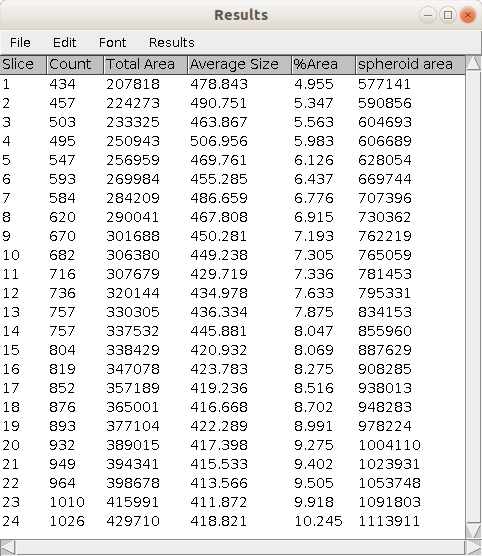
- Count: the number of detected nuclei in the frame (slice).
- Total Area: the total area of the detected nuclei in the frame (slice).
- Average Size: the average of the area of the nuclei in the frame (slice).
- spheroid area: the area of the spheroid in the frame (slice).
The image containing the rois as overlays and the results-table are saved into the results folder, the user specifies when running the tool.
How to cite the tool
You can use the tool's resource id (see scicrunch.org):
(Analyze Spheroid Cell Invasion In 3D Matrix, RRID:SCR_021204)
Publications using this tool
-
Menshikh, K., Antonaci, M., Radini, V., Salati, S., Daou, F., Cochis, A., Rimondini, L., 2025. Pulsed electromagnetic field stimulation subtly modulates doxorubicin sensitivity in a spheroid co-culture model of osteosarcoma. Sci Rep 15, 42441.
-
Kayley Jaworska, 2025. Developement of 3D in vitro models of glioblastoma (PhD). University of Huddersfield, Huddersfield, UK.
-
Quinteira, R., Gimondi, S., Monteiro, N.O., Sobreiro-Almeida, R., Lasagni, L., Romagnani, P., Neves, N.M., 2024. Decellularized kidney extracellular matrix-based hydrogels for renal tissue engineering. Acta Biomaterialia 180, 295–307. 10.1016/j.actbio.2024.04.026
-
Menshikh, K., Reddy, A.K., Cochis, A., Fraulini, F., Zambon, A., Lusvardi, G., Rimondini, L., 2024. Bifunctional mesoporous glasses for bone tissue engineering: Biological effects of doping with cerium and polyphenols in 2D and 3D in vitro models. Biomaterials and Biosystems 14, 100095. 10.1016/j.bbiosy.2024.100095
-
Moura, D.S., Mondaza-Hernandez, J.L., Sanchez-Bustos, P., Peña-Chilet, M., Cordero-Varela, J.A., Lopez-Alvarez, M., Carrillo-Garcia, J., Martin-Ruiz, M., Romero-Gonzalez, P., Renshaw-Calderon, M., Ramos, R., Marcilla, D., Alvarez-Alegret, R., Agra-Pujol, C., Izquierdo, F., Ortega-Medina, L., Martin-Davila, F., Hernandez-Leon, C.N., Romagosa, C., Salgado, M.A.V., Lavernia, J., Bagué, S., Mayodormo-Aranda, E., Alvarez, R., Valverde, C., Martinez-Trufero, J., Castilla-Ramirez, C., Gutierrez, A., Dopazo, J., Hindi, N., Garcia-Foncillas, J., Martin-Broto, J., 2024. HMGA1 regulates trabectedin sensitivity in advanced soft-tissue sarcoma (STS): A Spanish Group for Research on Sarcomas (GEIS) study. Cell. Mol. Life Sci. 81, 219. 10.1007/s00018-024-05250-y
-
Rodrigues, D.B., Moreira, H.R., Jarnalo, M., Horta, R., Marques, A.P., Reis, R.L., Pirraco, R.P., 2024. Generation of 3D melanoma models using an assembloid-based approach. Acta Biomaterialia 178, 93–110. 10.1016/j.actbio.2024.02.023
-
Bidan, N., Dunsmore, G., Ugrinic, M., Bied, M., Moreira, M., Deloménie, C., Ginhoux, F., Blériot, C., De La Fuente, M., and Mura, S. (2023). Multicellular tumor spheroid model to study the multifaceted role of tumor-associated macrophages in PDAC. Drug Deliv. and Transl. Res. 10.1007/s13346-023-01479-5.
-
Daniel Barreira Rodrigues, 2023. Bioengineering strategies for cancer therapy and modelling. University of Minho, Minho, Portugal.
-
Rikki Ann Mary Brown, 2023. Investigating the roles of microRNA-7-5p in head and neck squamous cell carcinoma and response to chemotherapy (PhD). University of Western Australia, Perth, Australia.
-
Anstee, J.E., Feehan, K.T., Opzoomer, J.W., Dean, I., Muller, H.P., Bahri, M., Cheung, T.S., Liakath-Ali, K., Liu, Z., Choy, D., et al. (2023). LYVE-1+ macrophages form a collaborative CCR5-dependent perivascular niche that influences chemotherapy responses in murine breast cancer. Developmental Cell, S1534580723003040. 10.1016/j.devcel.2023.06.006.
-
Mondaza-Hernandez, J.L., Moura, D.S., Lopez-Alvarez, M., Sanchez-Bustos, P., Blanco-Alcaina, E., Castilla-Ramirez, C., Collini, P., Merino-Garcia, J., Zamora, J., Carrillo-Garcia, J., et al. (2022). ISG15 as a prognostic biomarker in solitary fibrous tumour. Cell. Mol. Life Sci. 79, 434. 10.1007/s00018-022-04454-4.
-
Yang, H., Ting, X., Geng, Y.-H., Xie, Y., Nierenberg, J.L., Huo, Y.-F., Zhou, Y.-T., Huang, Y., Yu, Y.-Q., Yu, X.-Y., et al. (2022). The risk variant rs11836367 contributes to breast cancer onset and metastasis by attenuating Wnt signaling via regulating NTN4 expression. Sci. Adv. 8, eabn3509.
-
Hundsberger, H., Stierschneider, A., Sarne, V., Ripper, D., Schimon, J., Weitzenböck, H.P., Schild, D., Jacobi, N., Eger, A., Atzler, J., et al. (2021). Concentration-Dependent Pro- and Antitumor Activities of Quercetin in Human Melanoma Spheroids: Comparative Analysis of 2D and 3D Cell Culture Models. Molecules 26, 717.
-
Nousi, A., Søgaard, M.T., Audoin, M., and Jauffred, L. (2021). Single-cell tracking reveals super-spreading brain cancer cells with high persistence. Biochemistry and Biophysics Reports 28, 101120. 10.1016/j.bbrep.2021.101120.
-
Van Treuren, T., and Vishwanatha, J.K. (2018). CRISPR deletion of MIEN1 in breast cancer cells. PLoS ONE 13, e0204976.
-
Sperlich, J., and Teusch, N. (2018). Pseudopterosin Inhibits Proliferation and 3D Invasion in Triple-Negative Breast Cancer by Agonizing Glucocorticoid Receptor Alpha. Molecules 23, 1992.