Lipid Droplets Tool - MontpellierRessourcesImagerie/imagej_macros_and_scripts GitHub Wiki
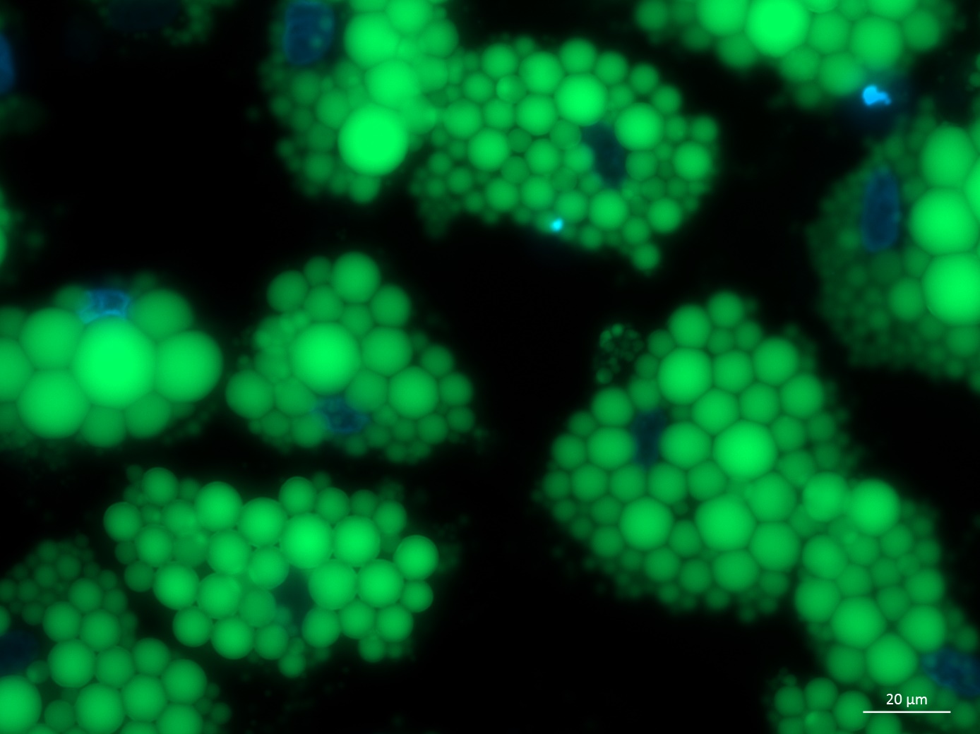
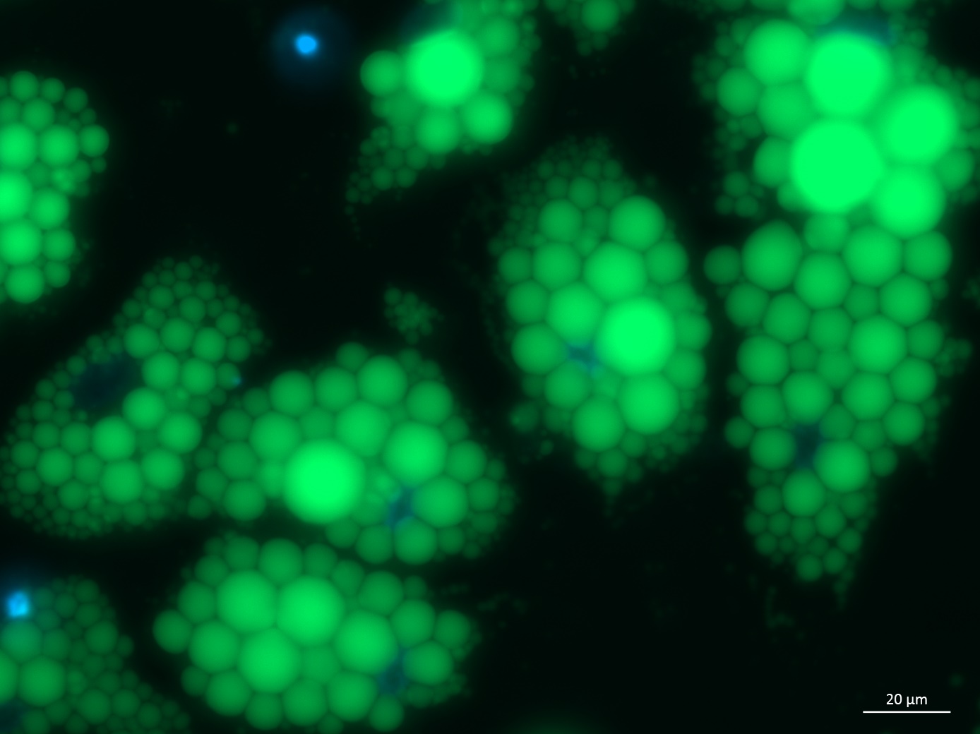
The Lipid Droplets Tool helps to segment lipid droplets marked with BODIPY.
You can find some example images here:
- Snap-8242%20SGBS%20BDNF10%2040x.jpg
- Snap-8235%20SGBS%20BDNF5%2040x.jpg
- Snap-8224%20SGGS%20BDNF1%2040x.jpg
Getting started
To install the tool, save the file MRI_Lipid_Droplets_Tool.ijm under macros/toolsets in the ImageJ installation and restart ImageJ.
Select the "MRI_Lipid_Droplets_Tool" toolset from the >> button of the ImageJ launcher.
- the first button (the one with the image) opens this help page
- the s button runs the segmentation on the current image
Options
Open the options-dialog by right-clicking on the s-button:
- min. size: objects smaller than min. size will be be considered as artefacts of the segmentation and will be removed from the result
Method
A bandpass filter is applied to the input image by using a Gaussian filter and then scaling the image down and up again with increasing scale and taking the difference between the two low-pass filtered images. On the result an automatic threshold (percentile method) is applied. Holes in the objects are filled. An auto-threshold (triangle) is applied to the input image and the resulting mask is used to remove artefacts from the mask of the droplets image. Objects smaller then a give size are removed and a binary watershed transform is used to separate touching droplets. Objects touching the border of the image are removed.
Publications using this tool
-
Pierre, L., Juszczak, F., Delmotte, V., Decarnoncle, M., Ledoux, B., Bultot, L., Bertrand, L., Boonen, M., Renard, P., Arnould, T., Declèves, A.-E., 2025. AMPK protects proximal tubular epithelial cells from lysosomal dysfunction and dedifferentiation induced by lipotoxicity. Autophagy 21, 860–880.
-
Morishige, J., Yoshioka, K., Nakata, H., Ishimaru, K., Nagata, N., Tanaka, T., Takuwa, Y., Ando, H., 2023. Sphingosine kinase 1 is involved in triglyceride breakdown by maintaining lysosomal integrity in brown adipocytes. Journal of Lipid Research 64, 100450.
-
Ali, U., Wabitsch, M., Tews, D., and Colitti, M. (2023). Effects of allicin on human Simpson-Golabi-Behmel syndrome cells in mediating browning phenotype. Front. Endocrinol. 14, 1141303. 10.3389/fendo.2023.1141303.
-
Florian Juszczak, 2022. New insights into therapeutic strategies for obesity-induced chronic kidney disease Is there a central role for AMP-activated protein kinase? (phD). University of Namur, Université de Mons.
-
Pessoa Rodrigues, C., Chatterjee, A., Wiese, M., Stehle, T., Szymanski, W., Shvedunova, M., Akhtar, A., 2021. Histone H4 lysine 16 acetylation controls central carbon metabolism and diet-induced obesity in mice. Nat Commun 12, 6212.
-
Hammel, J.H., and Bellas, E. (2020). Endothelial cell crosstalk improves browning but hinders white adipocyte maturation in 3D engineered adipose tissue. Integrative Biology 12, 81–89.
-
Montanari, T., Boschi, F., and Colitti, M. (2019). Comparison of the Effects of Browning-Inducing Capsaicin on Two Murine Adipocyte Models. Front. Physiol. 10, 1380.
-
Colitti, M., Boschi, F., and Montanari, T. (2018). Dynamic of lipid droplets and gene expression in response to β-aminoisobutyric acid treatment on 3T3-L1 cells. European Journal of Histochemistry 62.
-
Montanari, T., and Colitti, M. (2018). Simpson–Golabi–Behmel syndrome human adipocytes reveal a changing phenotype throughout differentiation. Histochemistry and Cell Biology, 149, 593–605.