Software Requirements Specification - CankayaUniversity/ceng-407-408-2019-2020-Image-Analysis-for-the-Classification-of-Brain-Tumor-Location-on-MR-Images GitHub Wiki
1. Introduction
I. Purpose
In past few decades, brain tumor segmentation in Magnetic Resonance Imaging (MRI) has become an emergent research area in the field of medical imaging systems. Accurate detection of brain tumor plays a important role in the diagnosis of brain cancer. Our project aims to develop a program which analyses the Magnetic Resonance Images of the patient under consideration and recognizes the tumor by using image processing algorithms and detects the location of tumor. By using this method, patients can get information about their results . Purpose of this document is to describe and give details about the project that planned to develop a method that detects brain tumor from MR Images. Research students, professors , teachers and all kind of people that wants to do exploration about ımage processing segmentation,artificial intelligence neural networks can get benefit from this document. This document will also be helpfull, for researchers who will do further projects about this area in the future and wants to improve the methods done so far.
II. Scope
This system involves determining the location and type of tumor in MRI images. The algorithms used today are not sufficient enough. On latest experiments, it is shown that hybrid method is one of the most successfull and proper method for image segmentation. We intend to use Hybrid Method to detect whether or not brain tumor exists in MR Image and to detect brain tumor region. If the algorithm shows the tumor region properly, we plan to use another Hybrid method to detect tumor type. We want to show whether the tumor is cancerous or not. This way, the doctors will have second hand verification system for brain tumors. Algorithms will detect and highlight the tumor region area. If detection of tumor area is sucessfull as planned, the method will pass on to second part of the project and will detect tumor type as cancerous or not. So, the second part of the project is optional. This proposed method/system will help the doctors in diagnosing brain tumors, and help the patients to start early medication/treatment.
III. Glossary
Medical Imaging: Medical imaging is the technique and process of creating visual representations of the interior of a body for clinical analysis and medical intervention, as well as visual representation of the function of some organs or tissues
Image processing: Image processing is a method to perform some operations on an image, in order to get an enhanced image or to extract some useful information from it. It is a type of signal processing in which input is an image and output may be image or characteristics/features associated with that image.
Image Segmentation: Image segmentation is the process of partitioning an image into multiple segments. Image segmentation is typically used to locate objects and boundaries in images.
Neural Networks: Neural networks are a set of algorithms, modeled loosely after the human brain, that are designed to recognize patterns.
Hybrid Method: Using more than one image processing algorithms on an image.
IV. References
[1] http://docwiki.embarcadero.com/RADStudio/Rio/en/Help_on_Help#Developer_Support_Services_and_Web_Site
[3] https://web.cs.dal.ca/~hawkey/3130/srs_template-ieee.doc
V. Overview
In the first part of this document we gave a brief description about the project and explained why this document is necessary. In the second part, we gave much more detail about the project, so it can be more understandable for readers and users of this method. Third part is much more detailed; this part gives software details of the project. Third part is mostly for users, researchers who wants to use this method on their own. Third part explains and gives helpful guide for them to use this method own their own. We gave detail about functional and non-functional features of this developed method.
2. Overall Description
I. Product Perspective:
The proposed method planned to work on both Linux and Windows operating systems. The method will work on brain MR Images only. The method will use the developed image processing algorithms already exists. We plan to use more than one image processing algorithms for detecting brain tumor area. We will use preprocessing for reducing noise on MR Images and use segmentation algorithms for detecting tumor area. The users of this method should have knowledge about medical structure of brain tumor or image segmentation or both. The code will be sharable and portable to the public after the method is published. The user can download the code on his/her computer and execute it for brain MR Images using any python editor.
II. User Characteristics:
The users of this method should have experience or knowledge about image segmentation and structure of brain and how it works. It is proposed that doctors in this area to use the method or research student and teachers who are interested in image processing and want to develop a project about this can do experiments with this method. It is recommended that doctors who use this method for their patients to not give the result of the method anyone other than their patients to prevent patient privacy. Users of this system should only use MR brain Images. User should know python programming language if they want to improve or test the proposed method.
III. Constraints:
The system only accepts Brain MR Images, if another image is given the system gives an error and does not work correctly. The system only works with specific operating systems. The system is coded in Python language. This code might not work with future version of python programming.
IV. Risks:
We did a research about medical image processing before starting this project. We plan to choose some of these articles we read and implement them, and combine the ones that gives the best result. The risk is that the implementation of these methods can not be fast enough so we may not start the second part of this project. Also, we will use different datasets from the experiments done in articles so the algorithms and efficiency might give different results depending on datasets. Finding enough amount of datasets and finding clean, accurate datasets are the biggest obstacles for this project
V. Assumptions and Dependencies
Future versions of python may not work with current version of python. The code of the method might have to change accordingly in the future.
3. Requirements
3.1 Specific Requirements
I. User Interfaces
There is no user interfaces for this Project since there is no website or application will be developed. The Project will be open source, and anyone who wants to access the code will be able to get phyton codes.
II. Hardware Interfaces
There is no hardware interface needed.
III. Software Interfaces
The codes will be available for both Linux and Windows. If the codes required for Linux and Windows Works differently,two different codes for both Linux and Windows will be shared.
IV. Communications Interfaces
No communication interface needed.
3.2 Functional Requirements
User should be able to see the original MR Image,ımage after segmentation that shows the tumor region and image after detection tumor type.
The tumor region and type should be understandable for the user.
The code should be easily readable and understandable for the user.
The system should detect tumor region accurately and should detect less than a minute.
The system should implement the algorithm properly and accordingly as; first preprocessing algorithms then segmentation algorithms.
3.3 Software system attributes
I. Reliability
The method will be published after, we make sure the algorithms are developed properly and the method is detection the area %100 accurately and fast enough as planned.
II. Avaibility
The method will be available for Linux and Windows operating systems. The code will be published and accessable for everyone to download.
III. Performance
The system should detect tumor region accurately and should detect less than a minute. The system should implement the algorithm properly and accordingly as; first preprocessing algorithms then segmentation algorithms.
IV. Portability
The system will be usefull in both Linux and Windows.
4.UML ANALYSIS MODEL
Use Case:
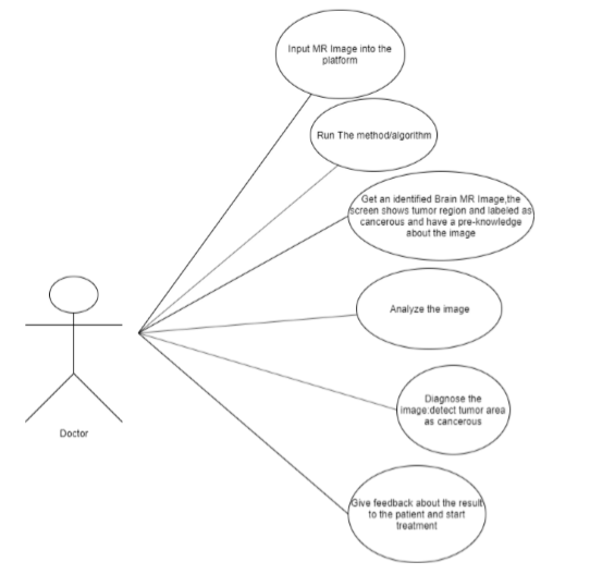
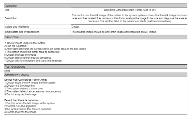
The following figure shows the use case diagram for the end users using the developed method. The most common users going to use the method will be doctors. This use case diagram shows the steps doctors can perform. Detecting Cancerous Tumor Region Use Case Diagram:
Detecting Cancerous Tumor Region Use Case Diagram:
Brief Description Of Use Case Diagram:
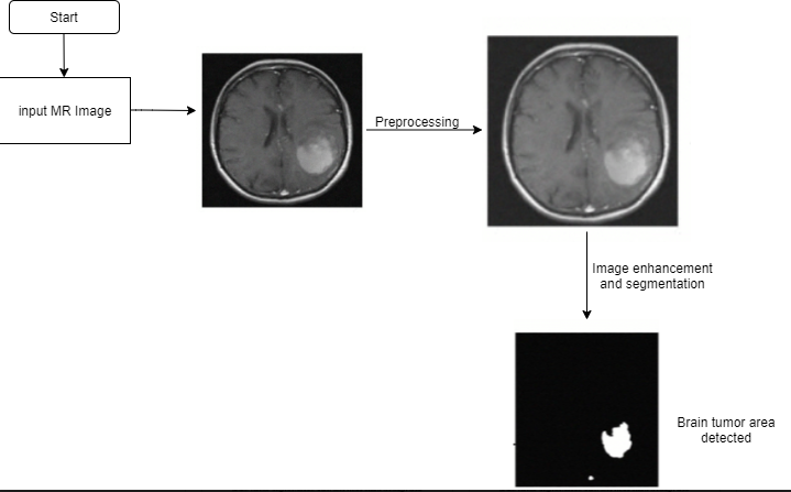
If a patient is suspected with a brain tumor and a brain MR Image of the patient is taken. Doctors can input the achieved MR Image into the system and run the algorithm developed from this project. After some time, the result of the algorithms and segmentation will be showed in the screen, this way doctor can have a pre-knowledge before doing diagnoses to the image, the developed methods can help and reduce time for doctor’s diagnoses. If there is no tumor found from the algorithms the screen won’t show any tumor area. If there is a tumor found from the algorithm; the screen will show the region of the tumor and identify it as non-cancerous or cancerous. The doctor will give the patient his/her result of the MR Image.
State Chart Diagram
State Chart of developed method:
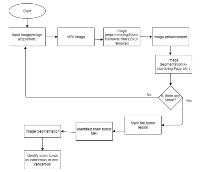
Flow chart is planned to be closer to this example: